What happens when your pet is sick or injured and your veterinarian needs to have a "closer look?" Sometimes, veterinarians need to use medical imaging to find out what's wrong with your furry friend or give him or her the best care possible.
That's normal enough, but do you know what medical imaging is, what options exist in veterinary care, or how medical imaging affects your dog or cat? Read on to learn more!
What is medical imaging?
Medical imaging is a diagnostic tool that allows veterinarians to take pictures of the inside of your pet in order to diagnose an ailment or ailments. A major benefit of medical imaging is that it is non-invasive, which means that no incision is necessary to produce an image of your pet.
Medical imaging is usually recommended when a veterinarian believes there is a problem with your pet that cannot be detected using a basic physical exam or blood test. There are four types of medical imaging available through veterinary medicine.

X-rays
X-rays, also known as radiographs, are the most common form of imaging used by veterinarians. Taking an x-ray involves exposing your pet to a beam of x-rays and taking a picture of their distribution as they pass through your pet. They are particularly useful for diagnosing fractures, arthritis, and pneumonia. However, not all diseases and conditions are apparent through x-rays, and for this reason your veterinarian may recommend other types of imaging .
As for the radiation, don’t worry: the amount of radiation your pet is exposed to during x-rays is minimal and harmless. If you see x-ray operators wearing protective gear, it is only because they are taking a precaution against accidental exposure to themselves.
Ultrasound
Imaging with sound waves is called ultrasound imaging, and is the second most common form of medical imaging in veterinary medicine. When an ultrasound examination is performed, a harmless, high-frequency sound beam – not detectable by humans or pets – is projected into the body of your pet. Ultrasound examinations are complementary to x-rays: they are especially useful in detecting abdominal diseases and are often able to provide a diagnosis when x-rays cannot.
CT and MRI Scans
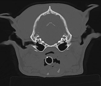
CT scanning, also known as “cat-scanning,” is a special type of x-ray exam in which a series of x-ray images, or “slices,” of your pet are obtained. CT scans are most useful when evaluating very complex parts of the body such as the head, chest, and some joints.
MRIs, by contrast, use a magnetic field and radio waves, rather than x-rays, to make images. MRIs can detect changes in body tissue by revealing increases in water and fluids due to inflammation or bleeding. MRIs are most useful in veterinary medicine to detect brain conditions such as strokes and spinal cord abnormalities such as herniated disks.

Will my pet need anesthesia or sedation?
This depends on how nervous or comfortable your pet is during the procedure, and to some degree on the type of imaging test performed.
For most x-ray procedures, no sedation or anesthesia is needed unless your pet is in pain and such options make your pet more comfortable. The same goes for ultrasound examinations.
On the other hand, anesthesia is almost always needed for CT and MRI examinations because it is very important that your pet remains still while images are being acquired. With some newer CT scanners, images are obtained very quickly, and this has allowed veterinarians and specialists to develop techniques to perform the tests with only sedation.
Does medical imaging always provide the final diagnosis?
That’s the goal, and occasionally it is possible to obtain a final answer from an imaging test. For example, x-rays might reveal a fracture as the cause of a limp or an ultrasound examination might clearly show a kidney stone.
However, many times the results of multiple tests are needed to determine a diagnosis. In fact, imaging tests often reveal the need for a totally different type of test, such as a biopsy. As a pet owner, you should be prepared for a logical progression of fact-finding, through multiple tests, to determine a final diagnosis of your pet’s ailment.
Getting to know your radiologist
Medical images are very complex and a veterinary radiologist may be needed to interpret the results accurately. Radiologists are licensed veterinarians who have completed 3-4 years of post-DVM training in diagnostic imaging interpretation and have passed a comprehensive certification examination.
If you have any questions or concerns, you should always visit or call your veterinarian – they are your best resource to ensure the health and well-being of your pets.
